Sunday, October 03, 2010

Make Typing Easy

By Bishnu Marasini
Sometimes we become irritated and tired when repeatedly typing some lengthy and/or bummer words (For example, we have to type, to get e.g.,) and symbols. Such case happens particularly during thesis, book, etc. writing. Scientific and mathematical symbols writing and their expression is more boring for some people (at least like me who hate typing comparing the other activities in front of computer). Although we might not have noticed, MS office has built facility to overcome this irritation. Just type eg (and space) to get e.g., just type temp (and space) to get temperature
  1. Click on MS office icon on the upper left corner.
  2. Click on word option. 
  3. Click/select on proofing
  4. Select AutoCorrect Option
  5. Select the AutoCorrect Tab
  6. (6A) Type the unique shortest form of the word for the desired word in left side and (6B) the desired (correct spelling) on the right side as shown in the figure.
  7. Click Replace to store on the database (on MS Word DLL) 
  8. Then OK.
  9. To insert frequently used symbol (like °C,α, β, etc.) select Math AutoCorrect tab.
  10. Don't forget to check on “Use Math AutoCorrect rules outside the math regions".
  11. Do/type again as described in 6 and as shown in the figure to get the desired result.
It will be only waste of time if this trick is done in those places (like cyber cafe) where OS is formatted in every week or month.

Friday, September 10, 2010

Switching Completely to Linux Operating System

By Bishnu Marasini 
       Although my engrossment in electronics as well as better understanding of computer hardware, I have not been working significantly in this field for 10 years (used to do at initial days). Only, as amateur technical support for my colleague is being the use and sharing of my expertise. While, parallel use of both Windows OS and Linux OS more than 10 years, I, now realized to switch completely towards Linux OS because of the following reasons.
  1. To Get rid of Virus: Windows XP is too old to resist virus attack, Windows Vista and Windows 7 also susceptible to attack, the pirated version are even worse. Linux OS might not have pulled hackers’ intention to enervate which may be due to fewer users and free/Open Source Software.
  2. Hardware Compatibility: The latest version of Microsoft like Windows Vista and Windows 7 cannot be installed or are very slow on the older CPU and Motherboard. The latest Linux OS always takes care of older CPU and motherboards but depends on the distro (type) of Linux.
  3. Better Understanding of Programming Code and Software Pleothera: Linux is free and Open Source Software and can be modified by anyone and redistribute the OS and we can read and copy every codes associated with it. This feature has been helping me to understand the meaning of code and my experiments in modifying only few terms from the code file and to see the ultimate effects. However, I have not developed any application.
  4. Better Security of Files/Pictures/Folders etc. I have very bad experience of corruption of my word (*.doc), excel (*.xls), pdf, pictures (*.jpg) while keeping them under XP OS. Which were damaged and decoded if could be opened. Fortunately, I have not lost them (except few due to backup duration/period cycle) because of monthly backup of data in Linux OS.
  5. Easy and Quicker Initial Installation of OS: Linux OS have the feature of complete installation along with all basic drivers e.g., driver of monitor, motherboard, audio drive, printer etc. in a single initial install. All of these drivers would have to be installed after the main Windows OS which ultimately increase installation time.
  6. Live CD: Because of live CD it made easier to understand, view features without any change and installation in hard disk. This also helps to restore/copy files and folder accidently deleted even in case of OS failure/crash.
  7. Swiftness: I have noticed that opening any application and internet search is very fast in Linux compared to Windows OS. Need for installation of anti-virus software and virus attack might impede the performance of Windows OS.
       Linux is free and becoming user friendly, but still more than 85% people pay to Microsoft/Apple Inc. for the same service because Linux OS still has many limitation.
  1. Difficult for Beginner of PC Users: Windows OS is easily understandable and could be one mouse click operation. Also, institute/schools use Windows OS to train basic computer, so they feel easy to use it in future days. However, some distros like Fedora, Ubuntu, OpenSuse, etc. have been progressing a lot in graphical user interface (GUI) so that it would be like Windows OS. They have improved in GNOME, KDE, Xfce, etc. desktop environment.
  2. Some Software and Games are not for Linux: Most of the daily required Software like MSOffice, ChemDraw, NamePro, SoftMax, ACD, Adobe Photoshop, Dreamweaver, etc. are only for Windows and can’t be installed in Linux. An adapter-like (emulator) software known as “Wine” facilitates to install these software in the Linux OS. But Wine has not exhibited satisfactory result in complete installation of most of the above applications. Also, it (Wine) opens the gate and vulnerability of virus to Linux root.
  3. Connection of Instruments to PC: Most instruments such as ELISA Reader, Microscope, PCR, etc., and even some printer can’t be connected to PC of Linux OS or if connected,  their full capacity enervated. A Distro “Scientific Linux” derived from Fedora and RHEL, "Scibuntu (UbuntuScience)" derived from Ubuntu may become better OS in future days not only to support such kind of scientific equipments but also better view of molecule’s, Protein’s, and DNA’s 3D structures in Linux OS.
  4. System Crash: As this OS is free and supported and built by community; some of its application and whole OS vulnerable to crash and should be debugged. The Distro “Debian” is found to be most stable but it is usually found to be outdated in comparison to other Linux Distro.
       At last, our smaller and smaller contribution like reporting of bug found in crash e.g., my report (https://bugzilla.redhat.com/show_bug.cgi?id=613592), sharing of scientific knowledge helps the volunteer engineer/developer to make Linux as better OS in future days. Also, we’ll not be accused of using pirated software used as lured by easiness and better performance; without paying in these days.
       I am currently using Fedora 12/13 as it is updated in every six months and latest repository and application of Linux is available via Fedora than other distro.

Wednesday, September 01, 2010

Identification of Beta-lactamase Inhibitor; a Strategy for Drug Development against Antibiotics Resistant Bacteria

Introduction:                                                                             By Bishnu Marasini

       The most threatening to human health is due to bacterial infection and intensive research has been addressing to treat these maladies since the known history. The infectious bacteria are microscopic, unicellular prokaryotes and different than mammalian eukaryotic cells. The outer layer of the bacterial cell consists of cell wall which is not found in mammalian cell and can be taken as target for development of bactericidal agents. The finding of clear zone of inhibition of bacterial culture around the growth of Penicillium notatum in the experiment of Alexander Fleming in 1928 was the positive indication to get rid of such threat.
       Nowadays almost all bacterial infection can be cured using antibiotics. The world consumes tons of antibiotics per year and half of them are beta-lactam type e.g. penicillin, amoxicillin, cephalosporin, cephalexin, cefixime, ceftriaxome, monobactam, carbapenem, methicillin etc. Beta-lactam antibiotics have broad spectrum activity, economical friendly on production, good safety profile, have clinical efficacy. It is also target specific for prokaryotic cells, little side effects except some allergic reaction. So, it has gained wide popularity.
       One of the components of bacterial cell wall is peptidoglycan which is cross linked polymer of repeatedly units of N-acetylglucosamine (NAG) and N-acetylmuramic acid (NAM). The final step in cell wall biosynthesis is transamidation reaction catalyzed by the enzyme cell wall transamidase (CWT) also called as penicillin binding protein (PBP). This enzyme helps to cross link the polymer of NAG and NAM. The highly strained and reactive beta-lactam ring of the antibiotics reacts and binds irreversibly with the serine hydroxyl group of PBP (shown in figure 1) which inactivates the enzyme and ultimately death of bacteria (Frère et al, 1984; Tipper and Strominger, 1965).

Figure1: Binding of β-lactam Ring with Penicillin Binding Protein
       However, beta-lactam antibiotics are becoming ineffective against pathogenic bacteria. The most common reason is due to the production of beta-lactamase enzyme (EC 3.5.2.6) which catalyze the hydrolysis of the antibiotics i.e., formation of carboxyl group degrading beta-lactam ring (Shown in figure 2). Hydrolyzed antibiotics lose its activity or binding affinity towards the PBP hence no effect against bacteria. Hydrolysis of beta-lactam is rapid by beta-lactamase than binding of beta-lactam to PBP (Bush 1988)

Figure 2: Degradation of β-lactam ring by β-lactamase enzyme
       The beta-lactamse (penicillinase) was reported just few years after the first antibiotic discovered (Abraham and Chain, 1940). Although penicillin is the oldest antibiotic and most of the organisms acquired resistant, it is first therapeutic choice in some diseases like syphilis. Beta-lactamse enzyme is an extra cellular enzyme in Gram-positive bacteria and found in periplasmic membrane in Gram-negative bacteria (Bowden and Georgiou, 1990; Dyke and Richmond, 1967). More than 200 types of beta-lactamse have been found (Bush et al, 1995). The difference among them is only the catalytic efficacy and turn over rate range from 0.004 to 1,200 molecules per second by 1 molecule of enzyme. Among them two types i.e. penicillinase and cephalosporinase type has a potent influence on the profile of the beta-lactam resistant antibiotics. Class A beta-lactamse has high affinity towards penicillin G but low affinity towards cephalosporin while class C beta-lactamse has opposite. Class B beta-lactamse hydrolyze the antibiotics by binding with the co-factor zinc (Zn) and class A, C and D hydrolyze by binding through serine residue of it to beta-lactam ring (Sawai et al, 1981). Beta-lactamase became widespread via the mechanism of plasmid exchange/insert among the pathogens (Sykes and Richmond, 1970). The rapid spread and evolution of these enzymes have seriously threatened the present antimicrobial arsenal.
       Two strategies have been developed to combat the problem of resistant. The first approach has been the synthesis/production of beta-lactamase resistant antibiotics e.g. penicillinase resistant beta-lactam antibiotics, nafcillin, oxacillin, ceftriaxone, cefoxitime, aztreonam, imipenem etc. But after few exposure to pathogens these antibiotics also become susceptible to extended spectrum beta-lactamase (ESBL) produced by multi-drug resistant (MDR) pathogens.
       The second approach is to use beta-lactamase inhibitors coupled with beta-lactam antibiotics. These enzyme inhibitors function to permanently inactivate the beta-lactamase in the periplasmic space so that the partner antibiotics can reach its target, penicillin binding protein (PBP). Broad spectrum beta-lactam antibiotics plus beta-lactamase inhibitors combination have been found good safety records and clinical efficacies (Munoz et al, 1996). Augmentin, the production of GlaxoSmithKline which is composed of amoxicillin and clavulanate in 2:1; Timentin (ticarcillin and clavulanate); Sultamicillin (ampicillin and sulbactam) are examples of beta-lactamase inhibitors in combination with beta-lactam used clinically. Clavulanate exhibited clinical efficacy than others and used as standard inhibitor. However, clavulanate was not found so effective against class C beta-lactamase (cephalorinase type) (Bush K, 1989). It also exhibited side effects on the long term of use like liver function destruction, gastrointestinal toxicity etc. (Ioannidis et al, 2002). Also it contains beta-lactam ring it and may be susceptible to beta-lactamase enzyme in upcoming days as broad spectrum beta-lactam antibiotics which were resistant to beta-lactamase, now become susceptible.

Tuesday, December 01, 2009

World AIDS Day

Recent Development in HIV Therapy (By Bishnu Marasini)
Human immunodeficiency virus (HIV), a lentivirus discovered in 1983 belongs to family Retroviridiae which is responsible for acquired immunodeficiency syndrome (AIDS), needs arduous work to eliminate. It contains two copies of +ssRNA which codes for 9 genes (~9 KB) is bound tightly to reverse transcriptase integrase and nucleocapsid proteins (p6 and p7), which protects RNA preventing digestion by nuclease. this nocleocapsid is surrounded by viral protein, p17 which also cover Vif, Vpr, Nef and viral protease. The envelope of virus contains some of the host cell membrane which is taken out during bulging out from infedted host cell. So, it contains glycoprotein gp120 and gp41 making envelope's spike. This spike structure is important to understand the replication cycle of the virus and drug development strategy (Schubert and Mcclure, 2005).
Due to error prone and rapid replication and hence immediate drug resistance, no successful single drug and neither immunization could be prepared against HIV. Although, some drugs are available are not sufficient to prevent epidemics, no better tolerated, convenient and cheap drugs are available. That’s why combination of drugs against different targets as well as development of new agents have been an exigent in recent days. Majority of these include reverse transcriptase, integrase, protease inhibitors, viral entry blockers, coating of CD4, CCR5, CXCR4, genetic therapy, immunotherapy (IL-7, IFN-α, IFN-γ) etc.

Reverse transcriptase (RT)
The two sub-units p66 (560 amino acids) and p51 (440 amino acids) together which make asymmetric heterodimer of RT. It acts as both DNA polymerage which copy RNA and as RNase H which degrades RNA only if the RNA is a part of an RNA/DNA duplex. This is very potent target for HIV therapy and nearly half of the discovered drug till date is based on its inhibition. Among them, Nucleoside Reverse Transcriptase Inhibitor (NRTI) and Non Nucleoside Reverse Transcriptase Inhibitor (NNRTI) have been catagorised. NRTIs are are structural analogues of the substrate of DNA lacking 3'-OH groups misleading to chain termination during replication. NRTI includes zidovudine, lamivudine, stavudine, didanosine, zalcifabine, abacavir, emtricitabine, tenofovir are already popular in HIV treatment. In addition, racivir, apricitabine, dexelvucitabine, elvucitabine, alovudine, amdoxovir etc. are in the current clinical development stage.
NNRITs are non-competitive inhibitors of RT binds to the pocket other than the active sites and change confirmation and/or inactivate the enzyme. NNRTI includes nevirapine, delavirdine (1st generation), efavirenz (2nd generation), rilpivirine. In addition, combination of these NRTI and NNRTIs has been widely searched for seeking of therapy e.g., Truvada (tenofovir/emtricitabine), Combivir (lamivudine and zidovudine), trizivir, dapivirine (Phase III).
RNAse H inhibitors include BBNH (N-(4-tert-Butylbenzoyl)-2-hydroxynaphthaldehyde hydrazone), DHBNH (dihydroxy benzoyl naphthyl hydrazone), CPHM (4-chlorophenylhydrazone of mesoxalic acid),  Diketo Acid( 4-[5-(Benzoylamino)thien-2-yl]-2,4-dioxobutanoic Acid), N-hydroxyimides, hydroxytropolone etc.

HIV-1 Protease Inhibitors
As already mentioned, HIV contains protease (aspertyl) which cleaves the group specific antigen (Gag) and Gag polymerage (Pol) precursor protein to structural capsid protein (p17, p24, p7 and p1) and functional protein (p11) reverse transcriptase (p66 and p51) and integrase (p32). All of these proteins activated after cleaving by viral protease, so, it has been a potent target for HIV-1 therapy. These includes saquinavir, totanavir, indinavir, nelfinavir, amprenavir, lopinavir, fosamprenavir etc. Side effects such as, lipodistrophy and dosing effect although seen in previous protease inhibitors, better one are atanazvir, brecanavir, darunavir, tipranavir, etc. are extensively under research and in the clinical trial phase which has not exhibited severe side effects as previous drugs.
HIV Integrase Inhibitors
HIV integrase transfer the viral genomic dsDNA into nucleus of host and insert into the host chromosome maintaining its stability, efficient expression and replication. If this enzyme is inhibited then replication and transcription of virus is blocked ultimately reducing viral load in the host body. Inhibitors of HIV integrase can be combined with other inhibitors for best effectiveness. These are efavirenz, raltegravir, elvitegravir etc.

HIV Maturation Inhibitors
At the late stage of the HIV proliferation, capsid precursor p25 (CA-SP1) is converted to mature capsid protein p24 (CA). This conversion can be blocked as HIV therapy. Bevirimat (3-O-(3',3'-dimethylsuccinyl)- betulinic acid) prevent the conversion of p25 to p24 capsid.
Viral Entry Inhibition
The outer spike of virus get attached to the T-cell and macrophase through the receptor and co-receptor (specially CD4, CCR5, CXCR4) present in the cell. Then nucleocapsid is integrated into host cells after attachment and fusion. The inhibition/blocking/coating of these receptor makes virus difficult to fuse with cell and entry. Mobozil, maraviroc, vicriviroc, aplaviroc etc. are the CCR5 and/or CXCR4 antagonist found as HIV therapeutics. However, aplaviroc found to be responsible in increasing liver enzyme and total billirubin. Enfuvirtide, a peptide inhibits the fusion of viral envelope with the host.
Gene Therapy
Although, most of the HIV infected person couldn't afford genetic therapy, significant HIV-infected patients live in setting where they not only afford but also contains sufficient infrastructure to support genetic therapy. Using of anti-sense, ribozymes, optamers, interference (RNAi), proteins (expressed), intrabodies, intrakin, zinc finger nuclease etc. are the the recent strategy for HIV therapy. Ribozymes against the gene tat, rev, and viral U5 has been in the clinical trial stage. Anti-sense RNA transgens pair with HIV RNA making non-functional duplex. Anti-U5 region anti-sensee RNA, envanti-sense RNA is being explored. Homology dependent control of gene activity is triggered by small-double stranded (sds) RNA molecules which is an RNAi regulatory mechanism. This has been applied in viral gene including gag, pol, nef, vif, env, vpr and LTR and showing exhibiting sign in cell line and potentially in HIV therapy in human beings. Another finding is the insertion of gene that express protein resembling mutant form of HIV Rev-protein (M10). It inhibits packing of virus and can be utilized by using retroviral or lentiviral vectors as HIV therapy.

Immunotherapy/ T-Cell Gene Therapy
It has been found that introducing CD28 co-stimulated T-cell reduce the viral infection. Also, genetic change in T-cells that express modified CD4+ significantly lower the virus load. In recent experiment CD4+ lymphocytes, genetically modified to express either protein Rev-M10 or a marking vector with no antiviral payload or both or to express an anti-sense TAR (Trans activating response region) element exhibited good antiviral effects. Gene expression pattern is designed such that it is associated with the acquisition of T-cell memory might be used to reprogram HIV specific T-cells to have TCM (long lived control memory T cell qualities. 

Others
Passive administration of interleukin (IL-2, IL-7) etc. and antibody specific against spike gp120 and gp41 of viral envelope, and other viral proteins. These not only eliminate/reduce virus load but also increase the number of CD4+ T-cells. IL-7 also protects CD4 and CD8 T-cells against apoptosis induced by HIV. Use of snake venom which contains phospholipase A2 (PLA2) protects human primary blood leukocytes from replication of HIV-1 strain, it also block viral entry into cells.
Conclusion
More than 25 drugs of HIV have been approved by FDA within the 20 years of time after first drug discovery. The combination of different specific target against HIV such as integrase inhibitors, reverse transcriptase inhibitors CCR5 inhibitors etc. can be administered as rescue therapy. Although the side effect, cost, availability, resistance acquired by virus is challenging the drug discovery realm. Research priority is being emphasized to vanquish this virus by human being. New better drugs of higher potency, less toxicity, lower risk of drug resistance development etc. are being developed in parallel with the shift point in calender, indicating good hope that the compliance of scientists but also politicians, Samaritans to stymie HIV.
References:
  1. Capitini CM, Chisti AA, Mackall CL (2009) Modulating T-cell homeostasis with IL-7: preclinical and clinical studies (Review), Journal of Internal Medicine, 266: 141-153.
  2. Luke W. Meredith, Haran Sivakumaran, Lee Major, Andreas Suhrbier, David Harrich (2009) Potent Inhibition of HIV-1 Replication by a Tat Mutant, PLoS ONE, 4 (11): e7769.
  3. Olga Latinovic, Janaki Kuruppu, Charles Davis, Nhut Le and Alonso Heredia (2009) Pharmacotherapy of HIV-1 Infection: Focus on CCR5 Antagonist Maraviroc (Review), Clinical Medicine: Therapeutics, 1: 1497-1510.
  4. Paul E. Sax, Camlin Tierney, Ann C. Collier et al, (2009) Abacavir–Lamivudine versus Tenofovir-Emtricitabine for Initial HIV-1 Therapy , New England Journal of Medicine, 361; 2230-2240.
  5. Stefan G. Sarafianos, Bruno Marchand, Kalyan Das, Daniel M. Himmel, Michael A. Parniak, Stephen H. Hughes and Eddy Arnold (2009) Structure and Function of HIV-1 Reverse Transcriptase: Molecular Mechanisms of Polymerization and Inhibition (Review), Journal of Molecular Biology, 385: 693-713.
  6. Ulirich Schubert and Myra Mcclure (2005), Topley and Wilson's Microbiology and Microbial Infection, Virology Vol 2, 10th edition, (Brian WJ Mahay and Volker Ter Meuleu: editors) Edward Arnold Ltd and ASM Press, page no. 1323-1345.
Bishnu Marasini

Saturday, June 20, 2009

परिवर्तन गर्नुपर्ने राजनैतीक थेगोहरु

नयाँ नेपाल ! डबल सेना पाल !!

जय नेपाल ! भए नेपाल !!

लाल सलाम ! पछाडि मन्त्रिको लाम !!

माओवादी जिन्दावाद ! खाओवादी खाएर पाद !!

नागरिक सर्वोच्चता ! रक्सिको भट्टी कता !!

पशुपतिनाथले सबैको कल्याण गरुन् ! बाराक ओबामाले पैसा भरुन् !!

Sunday, January 25, 2009

Issues on Animal/Human Cloning Difficulties and Challenges

Cloning:                                                                                                               By Bishnu Marasini
       The word cloning is derived from the Greek word ‘clone’ which means twig i.e. a group of identical entities. It is the process of making a genetically identical organism through non-sexual means (even growing a plant from cutting is a type of cloning). Most natural cloning occurs in those species which produce their descendents asexually e.g. bacteria, some fungi, Bermuda grass by means of runner stem etc. Animal cloning has been the subject of scientific experiment and the ‘Dolly’ is the first success. Here is only the cloning of human and animal is briefed.
             In 1885 Weishman put a theory stating that genetic information of a cell diminishes as the cell difference. In 1902, Hans S, split a two celled salamander embryo in two leading two adult salamanders this disproves Welshman’s hypothesis. In 1952 Robert B and Thomes JK cloned northern leopard frog using a method of nuclear transfer. A British biologist Handane JBS, in 1963 credited to have coined the term ‘clone’. Louis Jay Brown, the first child conceived through in vitro fertilization (IVF) (test-tube baby) was born in 25th July 1978. Ian W & Keith C in 23rd Feb. 1997 at Roslin Institute in Scotland announced the birth of ‘Dolly’, the first successful cloned animal. Again they created ‘Polly’ in June 1997 a lamb which was cloned from adult skin cells that was genetically altered to contain a human gene. Similarly, the birth of the first clone of an endangered animal, a baby bull ‘gaur’ named Noah was announced in 8th Jan 2001. The ‘Dolly’ was dead in 14th Feb 2003 due to progressive lung disease.

Method
i) Gaining of Oocytes/ egg cells: Various hormones like human menopausal gonadotropin (hMG), clomiphene, follicle stimulating hormone (FSH) is given for 5-10 days to the mature female of same species which is to be cloned.

ii) Enucleating of eggs/oocytes/somatic cell of donor: This is achieved by centrifuging cytochalastin-B treated cells such that nuclei detached from the eggs at the bottom of the tube leaving enucleated eggs in the supernatant. Cells/tissues taken from different organs like skin, bone, blood, mammary gland) of donor is cultured in petri-dish and single cell is isolated & nucleus is separated from cells which is ready to be injected in egg cells/oocytes.

iii) Injection of nucleus from donor (which is to be cloned)
  1. Using microinjection technique: The egg cells or oocytes is first immobilized by applying mild suction to the large blunt holding pipette & nucleus or DNA is then injected through the sharp end of a narrow glass micro needle.
  2. Isolated chromosomes with whole cells after co-precipitation with calcium phosphate.
  3. Fusion with electrical pulses.
iv) Culture: Thus the egg cells/oocytes injected with foreign nucleus (of donor) is cultured in vitro (2-8 cell stage) for certain period of time (48-72 hour) and then transferred into uterus.


v) Embryo transfer: embryo transfer in uterus of surrogate mother is done with the help of sonographer by using a Teflon catheter which is non sticky & done with 10 µl – 1 ml culture media. The surrogate mother is advised to rest for 24 hours and return in normal work after 48 hours.

Possible Uses:
  1. It is a good solution for increased infertility rate.
  2. To replace a child who has been lost by disease or accident.
  3. The duplication of individuals with particular talents or ability or importance.
  4. To conserve biodiversity including rare breeds.
  5. Non reproductive use of cloning (research cloning) is used for studying genetic change in cells derived from patients with conditions such as Alzheimer’s disease, Parkinson’s disease, diabetics etc.
  6. Making new animal models for human diseases, especially genetic diseases & for producing animals for use in xenotransplantation.
  7. The cloned animals will secret valuable recombinant protein and pharmaceuticals into their milk &/or blood &/or urine which can be used for extraction of these drugs; e.g. in sheep, ovine β-lactoglobulin gene promoter was fused in human a antitrypsin (hα,AT) gene which later produce hα,AT protein in milk used to treat the disease emphysema and cystic fibrosis.
Issues Difficulties and Challenges:
                 Ethical issue may arise regarding animals and human beings. There is no support in human cloning from the scientific community, religious community and other community and banned in various European countries and USA. If human cloning is allowed, only the rich may be benefited because of its cost. Also the surrogate mother may claim the child.
                 Success rate of cloning is very low, if succeed abnormal growth and/or disease may be seen because the cloned animal are vulnerable to be infected e.g. Dolly was suffered from arthritis and lung disease. Human cloning is more difficult than sheep or cattle because the cells of human embryos start producing proteins at relatively early stages. Cloning experiment in Japan show damage to immune systems, risks of death from pneumonia, liver failure, spontaneous abortion and abnormal births.
                Cloning procedure like research cloning seems to be very beneficial to human beings but their use and proper monitoring is essential and more and more people must be benefited.

Published in
Marasini, BP (2008), Microcosm, Vol. 3 No. 1

Thursday, May 08, 2008

Effect of Miotic/Mydriatic Agents on Eye and Molecular Mechanism

Eye:                                                                                        By Bishnu Marasini
        Eye maintains the proper visualization and clear image by controlling influx of light by smooth muscles that control the size of the pupil and the degree of adjustment is controlled by autonomic nervous system. These muscles are radial muscle of the iris (dilator papillae) (Iris dilator/radial muscle) and the circular muscle of the iris (constrictor papillae) (iris circular/sphincter muscle).
        Iris dilator/radial muscle is arranged radially in the iris and therefore fit as a dilator. It is innervated by the sympathetic system, which acts by releasing noradrenaline, which acts on α1-receptors. Its action to dilate lens let more light to reach in retina. Iris sphincter/circular muscle encircles the eye and constricts the lens. It is controlled by parasympathetic fibers that originate from the Edinger-Westphal nucleus, travel along the oculomotor nerve (CN III) through the superior orbital fissure and synapse in the ciliary ganglion, and then the postganglionic PSN fibers leave enter the eye via the short ciliary nerves. Acetylcholine is the transmitter of signal to it. The lens of the eye is attached to the cillary body by suspensory ligaments. When the circular/sphincter muscle is relaxed, the cillary body exerts tension on the lens causing it to flatten (for far vision). When the Circular/sphincter contracted; then lateral tension on the lens decreases and thickens (for near vision; cycloplegia). Circular and radial muscle act on the “tug of war” fashion; one is contracted other will relaxes.

Mydriasis and Miosis:
        “Mydriasis" is an excessive dilation of the pupil due to disease, trauma or the use of drugs. “Miosis” is constriction (or excessive, sometime abnormal) of the pupil of the eye.
       Mydriasis is mainly caused by the agents which are muscarinic (M3) antagonists (indirect) and adrenoreceptor (α1/α2) agonists (direct). Similarly miosis is caused by the agents which are muscarinic (M3) agonists (direct) and adrenoreceptor (α1/α2) antagonists (indirect). Also, cAMP inhibiting agents also reduce the degree of contraction of these muscles by decreasing Ca++ level in muscle by hyperpolarization. In addition to this; miotic/mydriatic agents might be systematically acting (administered in the part of body other than eye) or locally/topical acting (administered in the eye).
Miotic agents: dappiprazole (α1-antagonist), pilocarpine (M3 agonist), isoproterenol, thromboxane A2, yohimbine, Tolazoline, prostaglandin growth factor 2α (PGF2α), inomysin, thapsigargin etc.
Mydriatic agents: clonidine and tizanidine (α2 agonist), methoxamine (α1-agonist), phentolamine, Phenoxybenzamine, 8-chloroethylamine and phenylephrine (α1L-AR agonist), cyclopentalate and atropine (M3 antagonist), moxonidine, guanabenz etc. 

Mechanism:
        Pilocarpine increase the degree of contraction of sphincter muscle cell by activating Gq/11 signaling linked to M3 receptor. Atropine blocks activity of M3 agonist competitively binding on the receptor and sphincter muscle could not contract and dilator muscle vanquish on contraction as in tug of war fashion resulting mydriasis. 
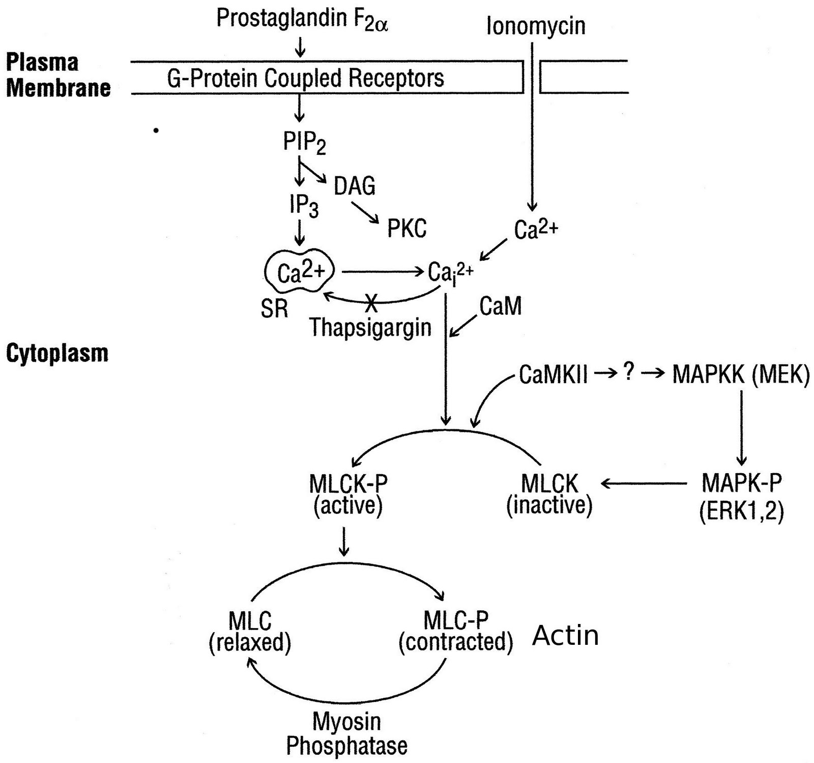
       Iris sphincter muscle cells express M3 receptor on their surface which is linked with pertussis toxin-insensitive G protein Gq/11, resulting in hydrolysis of membrane phospholipids; subsequent activation of phospholipase C-b (PLC-b) generates the major second messengers inositol (1,4,5)-trisphosphate (IP3) and diacylglycerol (DAG). Inositol(1,4,5)-trisphosphate binding to its receptor on intracellular storage sites results in mobilization/increasing cytosolic Ca2+ from extra cellular source and/or golgi bodies. Then Ca++ becomes available into the sarcoplasm and binds to troponin molecules in the thin filaments causing the troponoin to change the shape. This change in shape releases the troponin-tropomyosin complex from the myosin binding sites on the action. Contraction power strokes use ATP; myosin heads bind to actin, swivel and release; thin filaments are pulled toward center of sarcomere and finally contraction of the muscle. The increased Ca2+ in cytosol activates different mechanism/pathways; in which it actives rapid phosphorylation and activates of p42/p44 mitogen-activated Protein Kinase (MAPK), Ca2+/calmodulin-dependent protein kinase II (CaMKII) and myosin light chain kinase (MLCK), and other signaling cascade activating other enzymes as well as phosphorylation of myosin light chain (MLC) phosphorylation and contraction. 
      The miotic and mydriatic effect also found by nonadrenergic, noncholinergic response of the iris has been found in recent years in neurotransmitter binding site e.g., neurokinin A and B (NKA, NKB) which influx Ca2+ but not through the voltage-dependent channels, which are opened on depolarization of the membrane, mainly through the L and N types.
      The Ca2+ channels and the signalling pathways activated by ET-1 in the isolated rabbit iris sphincter. A scheme showing the signalling pathway activated by ET-1 to elevate [Ca2+]i and induce contraction is given below: We show that contraction of the iris sphincter by ET-1 or carbachol depends on the influx of extracellular Ca2+, as shown by abolition of the contraction after removal of extracellular Ca2+ nifedipine had minimal effects on the ET-1-induced functional responses. This latter observation is interesting because in cat iris sphincter, the Ca2+ mobilizing agonists PGF, ionomycin and thapsigargin increased MAP kinase phosphorylation and contraction.
      In the eye, the activation of EP1, EP4, and FP receptors by their selective agonists reduces intraocular pressure and causes pupil constriction.

Wednesday, March 26, 2008

Criteria for a Good Primer

By Bishnu Marasini
1. Primer Length:  It is important because melting temperature (Tm) and specificity partially relevant to primer length. Too long primer is inappropriate due to Tm and secondary structure formation and too short primer may not be specific. Generally 15-30 base long primer (or pair) is suitable.
2. Melting temperature (Tm): Tm of both primers (right and left primers that form a pair) is important while setting the annealing temperature in PCR reaction. Annealing temperature is usually set at 5°C less than the lower Tm (among the Tm of both primers). Generally the difference in Tm of both the primers also can’t be more than 5°C.
We can calculate the arbitrary Tm given by Wallace's rule for short length primer;
Tm = 4(G+C) + 2(A+T)°C
It should be around optimum Tm of template dsDNA and shouldn’t be deviate by more than 5°C.
3. %GC content: Although some genomic sequence have comparatively low GC content, around 50% GC content considered as suitable primer.
4. Self Annealing (SA): Self annealing of primer (forming like hair pin) make unavailable to anneal to template DNA. It can be scored as 1 (for each A-T pair) and 2 (for each G-C pair) when it forms the firmest hairpin structure and this score should be less than 20.
5. Pair Annealing (PA): Same as above (no. 4) when pair of primer anneals each other and it also should be less than 20.
6. Best Primer-pair Selection (total score of all of the above parameter): The final score of primer (or pair of primer) is calculated considering above mentioned parameter and best primer (or pair of primer) has the lowest value and always should be less than 10
score 1 per 10% difference in the optimal GC %
score 1 per °C difference from the optimal Tm
score 1 per 10 units of self annealing
score 1 per 10 units of pair annealing

NCBI’s online software “primer 3” for primer design (http://www.ncbi.nlm.nih.gov/tools/primer-blast/)

Saturday, December 01, 2007

Download for Desktop Background


You can download (Use as Desktop Background) and the image was taken fron Godawari Botanical Garden.

Wednesday, October 10, 2007

My Interest in Isolation and Identification of Escherichia coli from Urine Sample

The greenish shining colony in M-endo Agar distinguish E. coli from other Coliforms that help to separate E. coli from other bacteria present in stool and urine sample. Also, this method and media can be used to check contamination after antibacterial assay of E. coli. This method saved my time to identify by various sub-culture test.

Wednesday, October 03, 2007

Isolation and Identification of Salmonella spp. from Clinical/Food Sample

               The red colonies with black centers and media turn red (phenol red as indicator) in Xylose lysine deoxycholate (XLD) agar media distinguish Salmonella spp. from other coliforms that help to separate from other bacteria present in clinical blood/stool and food sample. Also, this method and media can be used to check contamination after antibacterial assay of Salmonella spp.
               Salmonellae can metabolize thiosulfate and produce hydrogen sulfide, hence black centers seen in colony but Shigellae can't and remains pink colony. Other coliforms are seen as yellow/orange colonies.
                Thus I separated Salmonella spp. very quickly from rest of the organisms.
 

Friday, September 21, 2007

Fare

We headed towards Koteswar from Balkhu, we were five talking about our honesty and innocuity. There was the conductor who had listened our expression. At destination, he asked for fare in greater rate, we discussed a while, but we gave the amount that he asked (greater than ususal).
Then one friend told that he had listened about our innocence, so he asked more fare and we could't resist.