Sunday, January 25, 2009

Issues on Animal/Human Cloning Difficulties and Challenges

Cloning:                                                                                                               By Bishnu Marasini
       The word cloning is derived from the Greek word ‘clone’ which means twig i.e. a group of identical entities. It is the process of making a genetically identical organism through non-sexual means (even growing a plant from cutting is a type of cloning). Most natural cloning occurs in those species which produce their descendents asexually e.g. bacteria, some fungi, Bermuda grass by means of runner stem etc. Animal cloning has been the subject of scientific experiment and the ‘Dolly’ is the first success. Here is only the cloning of human and animal is briefed.
             In 1885 Weishman put a theory stating that genetic information of a cell diminishes as the cell difference. In 1902, Hans S, split a two celled salamander embryo in two leading two adult salamanders this disproves Welshman’s hypothesis. In 1952 Robert B and Thomes JK cloned northern leopard frog using a method of nuclear transfer. A British biologist Handane JBS, in 1963 credited to have coined the term ‘clone’. Louis Jay Brown, the first child conceived through in vitro fertilization (IVF) (test-tube baby) was born in 25th July 1978. Ian W & Keith C in 23rd Feb. 1997 at Roslin Institute in Scotland announced the birth of ‘Dolly’, the first successful cloned animal. Again they created ‘Polly’ in June 1997 a lamb which was cloned from adult skin cells that was genetically altered to contain a human gene. Similarly, the birth of the first clone of an endangered animal, a baby bull ‘gaur’ named Noah was announced in 8th Jan 2001. The ‘Dolly’ was dead in 14th Feb 2003 due to progressive lung disease.

Method
i) Gaining of Oocytes/ egg cells: Various hormones like human menopausal gonadotropin (hMG), clomiphene, follicle stimulating hormone (FSH) is given for 5-10 days to the mature female of same species which is to be cloned.

ii) Enucleating of eggs/oocytes/somatic cell of donor: This is achieved by centrifuging cytochalastin-B treated cells such that nuclei detached from the eggs at the bottom of the tube leaving enucleated eggs in the supernatant. Cells/tissues taken from different organs like skin, bone, blood, mammary gland) of donor is cultured in petri-dish and single cell is isolated & nucleus is separated from cells which is ready to be injected in egg cells/oocytes.

iii) Injection of nucleus from donor (which is to be cloned)
  1. Using microinjection technique: The egg cells or oocytes is first immobilized by applying mild suction to the large blunt holding pipette & nucleus or DNA is then injected through the sharp end of a narrow glass micro needle.
  2. Isolated chromosomes with whole cells after co-precipitation with calcium phosphate.
  3. Fusion with electrical pulses.
iv) Culture: Thus the egg cells/oocytes injected with foreign nucleus (of donor) is cultured in vitro (2-8 cell stage) for certain period of time (48-72 hour) and then transferred into uterus.


v) Embryo transfer: embryo transfer in uterus of surrogate mother is done with the help of sonographer by using a Teflon catheter which is non sticky & done with 10 µl – 1 ml culture media. The surrogate mother is advised to rest for 24 hours and return in normal work after 48 hours.

Possible Uses:
  1. It is a good solution for increased infertility rate.
  2. To replace a child who has been lost by disease or accident.
  3. The duplication of individuals with particular talents or ability or importance.
  4. To conserve biodiversity including rare breeds.
  5. Non reproductive use of cloning (research cloning) is used for studying genetic change in cells derived from patients with conditions such as Alzheimer’s disease, Parkinson’s disease, diabetics etc.
  6. Making new animal models for human diseases, especially genetic diseases & for producing animals for use in xenotransplantation.
  7. The cloned animals will secret valuable recombinant protein and pharmaceuticals into their milk &/or blood &/or urine which can be used for extraction of these drugs; e.g. in sheep, ovine β-lactoglobulin gene promoter was fused in human a antitrypsin (hα,AT) gene which later produce hα,AT protein in milk used to treat the disease emphysema and cystic fibrosis.
Issues Difficulties and Challenges:
                 Ethical issue may arise regarding animals and human beings. There is no support in human cloning from the scientific community, religious community and other community and banned in various European countries and USA. If human cloning is allowed, only the rich may be benefited because of its cost. Also the surrogate mother may claim the child.
                 Success rate of cloning is very low, if succeed abnormal growth and/or disease may be seen because the cloned animal are vulnerable to be infected e.g. Dolly was suffered from arthritis and lung disease. Human cloning is more difficult than sheep or cattle because the cells of human embryos start producing proteins at relatively early stages. Cloning experiment in Japan show damage to immune systems, risks of death from pneumonia, liver failure, spontaneous abortion and abnormal births.
                Cloning procedure like research cloning seems to be very beneficial to human beings but their use and proper monitoring is essential and more and more people must be benefited.

Published in
Marasini, BP (2008), Microcosm, Vol. 3 No. 1

Thursday, May 08, 2008

Effect of Miotic/Mydriatic Agents on Eye and Molecular Mechanism

Eye:                                                                                        By Bishnu Marasini
        Eye maintains the proper visualization and clear image by controlling influx of light by smooth muscles that control the size of the pupil and the degree of adjustment is controlled by autonomic nervous system. These muscles are radial muscle of the iris (dilator papillae) (Iris dilator/radial muscle) and the circular muscle of the iris (constrictor papillae) (iris circular/sphincter muscle).
        Iris dilator/radial muscle is arranged radially in the iris and therefore fit as a dilator. It is innervated by the sympathetic system, which acts by releasing noradrenaline, which acts on α1-receptors. Its action to dilate lens let more light to reach in retina. Iris sphincter/circular muscle encircles the eye and constricts the lens. It is controlled by parasympathetic fibers that originate from the Edinger-Westphal nucleus, travel along the oculomotor nerve (CN III) through the superior orbital fissure and synapse in the ciliary ganglion, and then the postganglionic PSN fibers leave enter the eye via the short ciliary nerves. Acetylcholine is the transmitter of signal to it. The lens of the eye is attached to the cillary body by suspensory ligaments. When the circular/sphincter muscle is relaxed, the cillary body exerts tension on the lens causing it to flatten (for far vision). When the Circular/sphincter contracted; then lateral tension on the lens decreases and thickens (for near vision; cycloplegia). Circular and radial muscle act on the “tug of war” fashion; one is contracted other will relaxes.

Mydriasis and Miosis:
        “Mydriasis" is an excessive dilation of the pupil due to disease, trauma or the use of drugs. “Miosis” is constriction (or excessive, sometime abnormal) of the pupil of the eye.
       Mydriasis is mainly caused by the agents which are muscarinic (M3) antagonists (indirect) and adrenoreceptor (α1/α2) agonists (direct). Similarly miosis is caused by the agents which are muscarinic (M3) agonists (direct) and adrenoreceptor (α1/α2) antagonists (indirect). Also, cAMP inhibiting agents also reduce the degree of contraction of these muscles by decreasing Ca++ level in muscle by hyperpolarization. In addition to this; miotic/mydriatic agents might be systematically acting (administered in the part of body other than eye) or locally/topical acting (administered in the eye).
Miotic agents: dappiprazole (α1-antagonist), pilocarpine (M3 agonist), isoproterenol, thromboxane A2, yohimbine, Tolazoline, prostaglandin growth factor 2α (PGF2α), inomysin, thapsigargin etc.
Mydriatic agents: clonidine and tizanidine (α2 agonist), methoxamine (α1-agonist), phentolamine, Phenoxybenzamine, 8-chloroethylamine and phenylephrine (α1L-AR agonist), cyclopentalate and atropine (M3 antagonist), moxonidine, guanabenz etc. 

Mechanism:
        Pilocarpine increase the degree of contraction of sphincter muscle cell by activating Gq/11 signaling linked to M3 receptor. Atropine blocks activity of M3 agonist competitively binding on the receptor and sphincter muscle could not contract and dilator muscle vanquish on contraction as in tug of war fashion resulting mydriasis. 
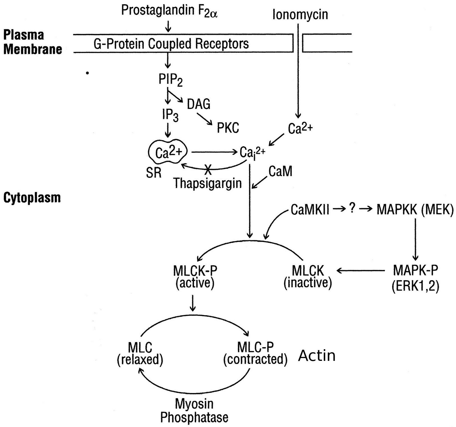
       Iris sphincter muscle cells express M3 receptor on their surface which is linked with pertussis toxin-insensitive G protein Gq/11, resulting in hydrolysis of membrane phospholipids; subsequent activation of phospholipase C-b (PLC-b) generates the major second messengers inositol (1,4,5)-trisphosphate (IP3) and diacylglycerol (DAG). Inositol(1,4,5)-trisphosphate binding to its receptor on intracellular storage sites results in mobilization/increasing cytosolic Ca2+ from extra cellular source and/or golgi bodies. Then Ca++ becomes available into the sarcoplasm and binds to troponin molecules in the thin filaments causing the troponoin to change the shape. This change in shape releases the troponin-tropomyosin complex from the myosin binding sites on the action. Contraction power strokes use ATP; myosin heads bind to actin, swivel and release; thin filaments are pulled toward center of sarcomere and finally contraction of the muscle. The increased Ca2+ in cytosol activates different mechanism/pathways; in which it actives rapid phosphorylation and activates of p42/p44 mitogen-activated Protein Kinase (MAPK), Ca2+/calmodulin-dependent protein kinase II (CaMKII) and myosin light chain kinase (MLCK), and other signaling cascade activating other enzymes as well as phosphorylation of myosin light chain (MLC) phosphorylation and contraction. 
      The miotic and mydriatic effect also found by nonadrenergic, noncholinergic response of the iris has been found in recent years in neurotransmitter binding site e.g., neurokinin A and B (NKA, NKB) which influx Ca2+ but not through the voltage-dependent channels, which are opened on depolarization of the membrane, mainly through the L and N types.
      The Ca2+ channels and the signalling pathways activated by ET-1 in the isolated rabbit iris sphincter. A scheme showing the signalling pathway activated by ET-1 to elevate [Ca2+]i and induce contraction is given below: We show that contraction of the iris sphincter by ET-1 or carbachol depends on the influx of extracellular Ca2+, as shown by abolition of the contraction after removal of extracellular Ca2+ nifedipine had minimal effects on the ET-1-induced functional responses. This latter observation is interesting because in cat iris sphincter, the Ca2+ mobilizing agonists PGF, ionomycin and thapsigargin increased MAP kinase phosphorylation and contraction.
      In the eye, the activation of EP1, EP4, and FP receptors by their selective agonists reduces intraocular pressure and causes pupil constriction.

Wednesday, March 26, 2008

Criteria for a Good Primer

By Bishnu Marasini
1. Primer Length:  It is important because melting temperature (Tm) and specificity partially relevant to primer length. Too long primer is inappropriate due to Tm and secondary structure formation and too short primer may not be specific. Generally 15-30 base long primer (or pair) is suitable.
2. Melting temperature (Tm): Tm of both primers (right and left primers that form a pair) is important while setting the annealing temperature in PCR reaction. Annealing temperature is usually set at 5°C less than the lower Tm (among the Tm of both primers). Generally the difference in Tm of both the primers also can’t be more than 5°C.
We can calculate the arbitrary Tm given by Wallace's rule for short length primer;
Tm = 4(G+C) + 2(A+T)°C
It should be around optimum Tm of template dsDNA and shouldn’t be deviate by more than 5°C.
3. %GC content: Although some genomic sequence have comparatively low GC content, around 50% GC content considered as suitable primer.
4. Self Annealing (SA): Self annealing of primer (forming like hair pin) make unavailable to anneal to template DNA. It can be scored as 1 (for each A-T pair) and 2 (for each G-C pair) when it forms the firmest hairpin structure and this score should be less than 20.
5. Pair Annealing (PA): Same as above (no. 4) when pair of primer anneals each other and it also should be less than 20.
6. Best Primer-pair Selection (total score of all of the above parameter): The final score of primer (or pair of primer) is calculated considering above mentioned parameter and best primer (or pair of primer) has the lowest value and always should be less than 10
score 1 per 10% difference in the optimal GC %
score 1 per °C difference from the optimal Tm
score 1 per 10 units of self annealing
score 1 per 10 units of pair annealing

NCBI’s online software “primer 3” for primer design (http://www.ncbi.nlm.nih.gov/tools/primer-blast/)

Saturday, December 01, 2007

Download for Desktop Background


You can download (Use as Desktop Background) and the image was taken fron Godawari Botanical Garden.

Wednesday, October 10, 2007

My Interest in Isolation and Identification of Escherichia coli from Urine Sample

The greenish shining colony in M-endo Agar distinguish E. coli from other Coliforms that help to separate E. coli from other bacteria present in stool and urine sample. Also, this method and media can be used to check contamination after antibacterial assay of E. coli. This method saved my time to identify by various sub-culture test.

Wednesday, October 03, 2007

Isolation and Identification of Salmonella spp. from Clinical/Food Sample

               The red colonies with black centers and media turn red (phenol red as indicator) in Xylose lysine deoxycholate (XLD) agar media distinguish Salmonella spp. from other coliforms that help to separate from other bacteria present in clinical blood/stool and food sample. Also, this method and media can be used to check contamination after antibacterial assay of Salmonella spp.
               Salmonellae can metabolize thiosulfate and produce hydrogen sulfide, hence black centers seen in colony but Shigellae can't and remains pink colony. Other coliforms are seen as yellow/orange colonies.
                Thus I separated Salmonella spp. very quickly from rest of the organisms.
 

Friday, September 21, 2007

Fare

We headed towards Koteswar from Balkhu, we were five talking about our honesty and innocuity. There was the conductor who had listened our expression. At destination, he asked for fare in greater rate, we discussed a while, but we gave the amount that he asked (greater than ususal).
Then one friend told that he had listened about our innocence, so he asked more fare and we could't resist.

Wednesday, August 22, 2007

jokes

Quiet in Church
A Sunday school teacher asked the children just before she dismissed them to go to church,

"And why is it necessary to be quiet in church?"

Little Birkhe jumped up and yelled, "Because people are sleeping!"


The New Baby
Little Birkhe 's next door neighbors had a baby. Unfortunatly, the little baby was born with no ears. When they arrived home from the hospital, the parents invited Little Birkhe's family to come over and see their new baby.

Little Birkhe 's parents were very afraid that their son would have a wise crack to say about the baby so the dad had a long talk with little Birkhe before going to the neighbors.

He said "Now, son... that poor baby was born without any ears. I want you to be on your best behavior and not say one word about his ears or I am really going to spank you when we get back home."

"I promise not to mention his ears at all" said Little Birkhe.

At the neighbor’s home, Little Birkhe leaned over in the crib and touched the baby's hand. He looked at it's mother and said "Oh what a beautiful little baby".

The mother said "Thank you very much, Little Birkhe."

He then said, "this baby has perfect little hands and perfect little feet. Why... just look at his pretty little eyes.... Did his doctor say that he can see good?"

The Mother said "why, yes... his doctor said he has 20/20 vision.

Little Birkhe said "well, it is a darn good thing cause he sure couldn't wear glasses!!!


Two Fingers
It's the first day of school and the teacher told her kindergarten class, "If anyone has to go to the toilet, you should hold up two fingers."

After a moment of quiet thought, Little Birkhe asked: "How will that help?"

Wednesday, May 23, 2007

Involvement of protein kinase C- in DNA damage-induced apoptosis

Caspases are essential for the execution of cell death by apoptotic stimuli.1,2,3,4 The pathway of cell death varies depending on the cell type as well as the apoptotic stimuli. It is generally believed that binding of Fas ligand or tumor necrosis factor- (TNF) to their receptors causes activation of the initiator caspase-8 followed by the activation of a caspase cascade to execute cell death.2,5 In contrast, DNA damaging agents are known to induce release of mitochondrial Cytochrome c, which facilitates the interaction of apoptotic protease activating factor (Apaf-1) with procaspase-9 to initiate the activation of downstream effector caspases, such as caspases-3 or -7 to cause cell death.3 Both receptor-mediated and anticancer drug-induced apoptosis may, however, involve more than one pathway and there may be cross-talk between these two pathways.6,7,8

cis-Diamminedichloroplatinum(II) (cDDP or cisplatin) is one of the most important anticancer agents used for the treatment of solid tumors.9 Although the antitumor activity of cDDP is believed to be due to its interaction with chromosomal DNA, only a small fraction of cDDP actually interacts with DNA and inhibition of DNA replication cannot solely account for its biological activity.10 The efficacy of chemotherapeutic drugs not only depends on their ability to induce DNA damage but also on the cell's ability to detect and respond to DNA damage.11 cDDP, like other chemotherapeutic drugs, causes activation of caspases although the sequence of events that follow cDDP-induced DNA damage and lead to apoptosis remains to be unraveled.

We and others have shown that the PKC signal transduction pathway regulates cell death by cDDP.12,13,14,15,16,17 PKC is a family of 11 isozymes that are classified as the conventional PKCs (, I, II and ), novel PKCs (, , , and ), atypical PKCs ( and / ) and novel/atypical PKC.18 PKC is a substrate for caspase-3 and the catalytic fragment of PKC has been directly associated with apoptotic cell death.19,20 We have, however, demonstrated that downregulation of PKC that decreased the abundance of PKC catalytic fragment was associated with increased cellular sensitivity to cDDP.17 These results raise the possibility that PKC acts upstream of caspases to regulate cell death by cDDP. It is not known which PKC isozyme regulates activation of caspases and which step(s) of the cisplatin-induced cell death pathway is regulated by PKC.

Mitochondria play a pivotal role in the decision making process of a cell's life and death.21 It is believed that once Cytochrome c is released from mitochondria, cells are committed to die.22 An inability to induce release of Cytochrome c from mitochondria has been associated with cellular resistance to anticancer agents, including cDDP.23 In the present study, we have investigated how PKC regulates release of Cytochrome c and activation of caspases that emanate from mitochondria. Our results show that in HeLa cells, PKC was localized not only in the cytosol but also in the mitochondrial fraction and cDDP induced processing of both cytosolic and membrane-associated PKC. Rottlerin, a specific inhibitor of PKC, blocked cDDP-induced activation of caspases and proteolytic cleavage of PKC in both cytosolic and HM fractions but inhibited only late but not early release of Cytochrome c. Furthermore, inhibition of nPKC, but not of cPKCs, protected cells against cDDP-induced cell death. Taken together, these results demonstrate that PKC influences cDDP-induced cell death by acting at an early step of the mitochondrial cell death pathway that precedes activation of caspases.

Friday, May 04, 2007

Camphor

Description
The camphor tree is a dense broadleaved evergreen that is capable of growing 50-150 ft (15.2-45.7 m) tall and spreading twice that wide with a trunk up to 15 ft (4.6 m) in diameter, though the largest U.S. specimens are only half that size and those in the Caribbean are even smaller. The shiny foliage is made up of alternate 1-4 in (2.5-10.2 cm) oval leaves dangling from long petioles. Each leaf has three distinct yellowish veins. The outer margins of the leaves tend to be somewhat wavy and turn upward. The new foliage starts out a rusty burgundy color, but the leaves soon turn dark green on the upper sides and paler green underneath. New branches emerging from the shallowly fissured grayish brown trunk are smooth and green. Twigs are usually green, but may be tinged with red when young. The inconspicuous tiny cream colored flowers are borne in the spring on branching 3 in (7.6 cm) flower stalks. They are followed by large crops of fruit, comprised of round pea sized berries attached to the branchlets by cuplike little green cones. The berries first turn reddish, then ripen to black. Camphor tree can be readily identified by the distinctive odor of a crushed leaf.


This camphor tree shades a house in North Florida - a landscape use no longer considered appropriate for this invasive species.
Location
Cinnamomum camphora, the camphor tree, comes from China, Japan, Korea, Taiwan, and adjacent parts of East Asia, where it grows in mesic forests and on well-drained sites along streambanks. Camphor has become widely naturalized in Australia. In the United States, it is grown along the Gulf Coast and in California, and has escaped cultivation and become naturalized in many areas.

Culture
Camphor prefers fertile sandy soil. It will tolerate a pH anywhere in the range of 4.3 to 8. The roots are very sensitive to disturbance. They may extend far from the trunk of the tree, and can readily be identified by their characteristic odor.
Light: Camphor will grow in full sun or partial shade.
Moisture: Camphor tree does not do well in wet soils. Established trees are tolerant of drought.
Hardiness: USDA Zones 8 - 10. Hardened off camphor trees can survive freezes down to 10-15ºF (-12 - -9ºC), but new growth will suffer freeze burn when the temperature drops below 32ºF (0ºC) and branches will die back from temperatures in the low twenties.
Propagation: Camphor seed does not remain viable for long and should be planted in the greenhouse as soon as it ripens. Remove the fruit pulp first. At 68ºF (20ºC), germination will take 1-6 months. Cuttings of semiripe side shoots can be rooted in a warm humid place in midsummer. Pieces 2-3 in (5.1-7.6 cm) long with a heel work best.

Usage
Camphor is widely planted as a shade tree, screen, or windbreak. In China and Japan, it is grown commercially for its medicinal oil. Camphor oil has a strong penetrating fragrance, a pungent bitter flavor, and feels cool on the skin like menthol, though it also has irritating qualities as well as a numbing effect. Camphor has been used to treat ailments ranging from parasitic infections to toothaches. Scientific evidence has confirmed that chemicals in the plant have value in antiseptics and medications for treating diarrhea, inflammation, itching, and nervous conditions. Camphor wood is prized for its attractive red and yellow striping, amenability to woodworking, and insect repelling properties. It is light to medium in weight and soft to medium in hardness. Wood from the camphor tree is not especially strong, but it takes polishing well. It is commonly used for chests, closets, coffins, instruments, and sculptures. Camphor veneer is used in fine cabinetry. Camphor is also used in perfumes.


camphor tree trunk and bark
Features
This is a sturdy storm resistant tree which makes a good windbreak. Since it is hard to burn, it should also be valuable as a shade tree in areas that are prone to wildfires. Unfortunately, these desirable traits are offset by the tree's invasiveness and damaging effects on wildlife and natural communities. This fine tree should be grown and appreciated in its native range, but not planted in other regions where species and ecosystems have not adapted to its aggressiveness and toxicity. Camphor tree should not be grown in the United States.

WARNING
Camphor in large doses is toxic to humans. It stimulates the central nervous system and may affect respiration or cause convulsions. In Chinese medicine, camphor is forbidden for pregnant women and those with a deficiency of vital energy or yin. Camphor is a prolific seed producer that apparently does not have serious predators or diseases outside its native range. Seedlings and root sprouts are abundant near mature trees, but individual trees pop up far from seed sources. In Florida, camphor trees appear in undisturbed mesic hardwood forests, upland pine woods, and scrubs, as well as in the vacant lots and fencerows where it is more commonly observed. The Plant Conservation Alliance lists this species as an Alien Invader and it is listed as a Category I invasive exotic species by the Florida Exotic Pest Plant Council, which means that it is known to be "invading and disrupting native plant communities in Florida

Saturday, March 10, 2007

chyura

When we had gone for a dry picnic, there was "chyura" for us. My friend told me, "I don't like chyura and I know you like most the chyura." I wander and asked how did you know that but I also don't like chyura. He told me, "I always see that lots of packets of chyura when I meet your room." I smiled, wrong interpretation had you ever seen finished that?

Wednesday, March 07, 2007

Holi

My brother asked, "Holi kasto bhayo?"
(Because I get up at 8-9 AM) I replied "Uttthne bittikai holi kheleko Jado bhayo"